Holmium »
PDB 1buu-9c90 »
1rer »
Holmium in PDB 1rer: Crystal Structure of the Homotrimer of Fusion Glycoprotein E1 From Semliki Forest Virus.
Protein crystallography data
The structure of Crystal Structure of the Homotrimer of Fusion Glycoprotein E1 From Semliki Forest Virus., PDB code: 1rer
was solved by
D.L.Gibbons,
M.C.Vaney,
A.Roussel,
A.Vigouroux,
B.Reilly,
M.Kielian,
F.A.Rey,
with X-Ray Crystallography technique. A brief refinement statistics is given in the table below:
| Resolution Low / High (Å) | 20.00 / 3.20 |
| Space group | P 31 2 1 |
| Cell size a, b, c (Å), α, β, γ (°) | 198.197, 198.197, 116.250, 90.00, 90.00, 120.00 |
| R / Rfree (%) | 26.5 / 28.5 |
Other elements in 1rer:
The structure of Crystal Structure of the Homotrimer of Fusion Glycoprotein E1 From Semliki Forest Virus. also contains other interesting chemical elements:
| Bromine | (Br) | 3 atoms |
Holmium Binding Sites:
The binding sites of Holmium atom in the Crystal Structure of the Homotrimer of Fusion Glycoprotein E1 From Semliki Forest Virus.
(pdb code 1rer). This binding sites where shown within
5.0 Angstroms radius around Holmium atom.
In total 4 binding sites of Holmium where determined in the Crystal Structure of the Homotrimer of Fusion Glycoprotein E1 From Semliki Forest Virus., PDB code: 1rer:
Jump to Holmium binding site number: 1; 2; 3; 4;
In total 4 binding sites of Holmium where determined in the Crystal Structure of the Homotrimer of Fusion Glycoprotein E1 From Semliki Forest Virus., PDB code: 1rer:
Jump to Holmium binding site number: 1; 2; 3; 4;
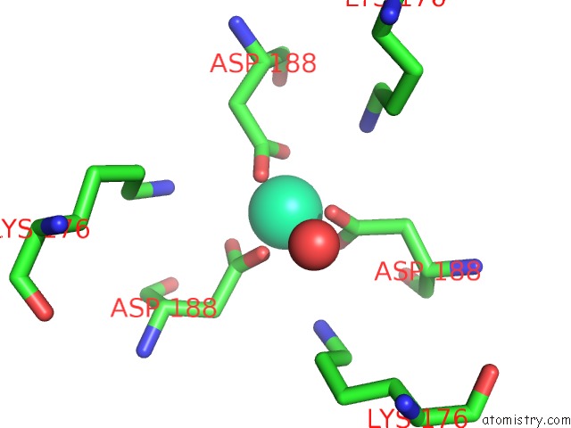
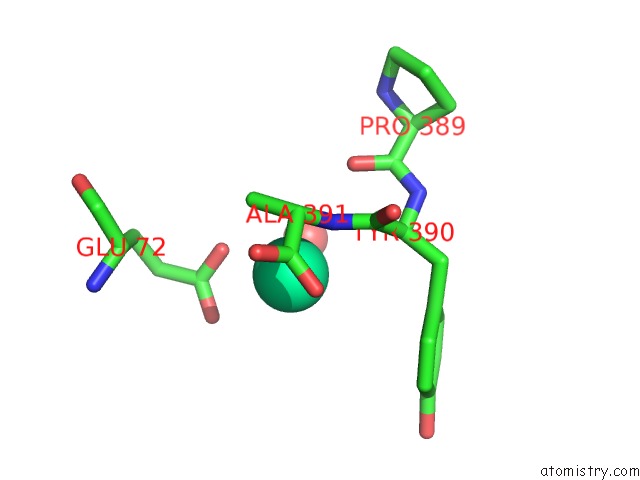
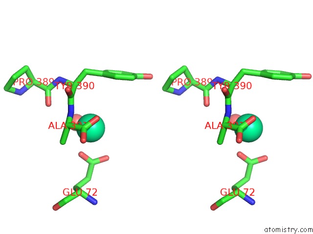
Holmium binding site 1 out of 4 in 1rer
Go back to
Holmium binding site 1 out
of 4 in the Crystal Structure of the Homotrimer of Fusion Glycoprotein E1 From Semliki Forest Virus.
Mono view
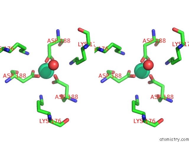
Stereo pair view
Mono view
Stereo pair view
A full contact list of Holmium with other atoms in the Ho binding
site number 1 of Crystal Structure of the Homotrimer of Fusion Glycoprotein E1 From Semliki Forest Virus. within 5.0Å range:
|
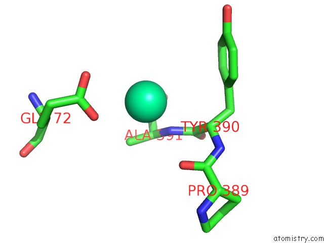
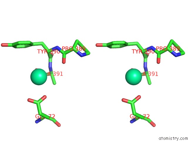
Holmium binding site 2 out of 4 in 1rer
Go back to
Holmium binding site 2 out
of 4 in the Crystal Structure of the Homotrimer of Fusion Glycoprotein E1 From Semliki Forest Virus.
Mono view
Stereo pair view
Mono view
Stereo pair view
A full contact list of Holmium with other atoms in the Ho binding
site number 2 of Crystal Structure of the Homotrimer of Fusion Glycoprotein E1 From Semliki Forest Virus. within 5.0Å range:
|
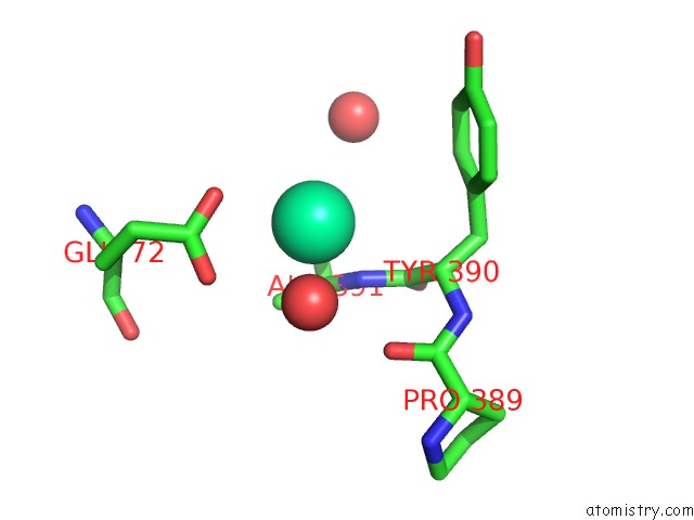
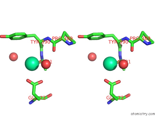
Holmium binding site 3 out of 4 in 1rer
Go back to
Holmium binding site 3 out
of 4 in the Crystal Structure of the Homotrimer of Fusion Glycoprotein E1 From Semliki Forest Virus.
Mono view
Stereo pair view
Mono view
Stereo pair view
A full contact list of Holmium with other atoms in the Ho binding
site number 3 of Crystal Structure of the Homotrimer of Fusion Glycoprotein E1 From Semliki Forest Virus. within 5.0Å range:
|
Holmium binding site 4 out of 4 in 1rer
Go back to
Holmium binding site 4 out
of 4 in the Crystal Structure of the Homotrimer of Fusion Glycoprotein E1 From Semliki Forest Virus.
Mono view
Stereo pair view
Mono view
Stereo pair view
A full contact list of Holmium with other atoms in the Ho binding
site number 4 of Crystal Structure of the Homotrimer of Fusion Glycoprotein E1 From Semliki Forest Virus. within 5.0Å range:
|
Reference:
D.L.Gibbons,
M.C.Vaney,
A.Roussel,
A.Vigouroux,
B.Reilly,
J.Lepault,
M.Kielian,
F.A.Rey.
Conformational Change and Protein-Protein Interactions of the Fusion Protein of Semliki Forest Virus. Nature V. 427 320 2004.
ISSN: ISSN 0028-0836
PubMed: 14737160
DOI: 10.1038/NATURE02239
Page generated: Sun Aug 11 09:14:18 2024
ISSN: ISSN 0028-0836
PubMed: 14737160
DOI: 10.1038/NATURE02239
Last articles
Zn in 9MJ5Zn in 9HNW
Zn in 9G0L
Zn in 9FNE
Zn in 9DZN
Zn in 9E0I
Zn in 9D32
Zn in 9DAK
Zn in 8ZXC
Zn in 8ZUF